Effect of Human Cryopreservation Protocol on the Ultrastructure of the Canine Brain (Platt)
TABLE OF CONTENTS
Introduction
Brief lay summary of results
Summarized extracts from the paper
Electron micrographs
Original technical paper by Darwin et. al. (on different web page)
The electron micrographs on this page are also available in a high resolution PDF file for production of hardcopies. Right Click the link and choose “Save Target As” to download this 5-megabyte file.
Introduction: New Brain Study Shows Reduced Tissue Damage
From CryoCare Report #4
Online Edition, July 1995
by Charles Platt
New evidence shows that when human cryopreservation is carried out under favorable conditions, it causes minimal damage that we can reasonably expect to be reversible at some time in the future using molecular nanotechnology.
A month ago, I would not have been able to write that sentence. I would have felt compelled to qualify it in some way – because no study of human cryopreservation protocol had ever shown freezing damage that could genuinely be described as “minimal.”
Now, however, research by Michael Darwin, Sandra Russell, Paul Wakfer, Larry Wood, Candy Wood, and Steven B. Harris MD has given us new reason to believe that modern cryopreservation techniques are doing what they’re supposed to do: minimizing freezing damage more successfully than simpler perfusion protocols that were used in the past.
First let me recap some basic background. In the 1950s, experiments showed that when the cells of a mammal are frozen, they experience less damage if they have been soaked previously in a solution of glycerol.
Unfortunately, it’s much more difficult to apply this treatment to an intact organism than to a small tissue sample. Glycerol is capable of causing excessive tissue shrinkage and damage to cells if it’s administered too rapidly in too high a concentration. Using proper introduction and monitoring equipment, however, the concentration of the solution can be gradually ramped up during a prolonged period of perfusion so that a high terminal concentration can be reached with relative safety. This technique was applied to cryonics patients by Leaf, Darwin, and others during the 1980s.
While there were good reasons to believe that this protocol was giving patients an improved level of protection, the supposition was never properly verified, mainly because there was insufficient time, money, and personnel to support the research. But in 1993, when BioPreservation moved into its new home in the building owned by Twenty-First Century Medicine, there was an opportunity to catch up on this overdue verification.
Several dogs were anesthetized and were put into cardiac arrest while they were unconscious. After a short waiting period (equivalent to the wait that a cryonics patient might experience before receiving attention from a transport team), the dogs were given cardio-pulmonary support using a “Thumper” mechanical CPR device, medications were administered, and blood washout and perfusion with glycerol were carried out, in exactly the same way as if the BioPreservation team was dealing with a human cryonics case. The dogs were cooled and maintained for 12 to 18 months at -90 C, then rewarmed. Brain samples were examined using light and electron microscopy, and damage was found to be minimal.
A second set of dogs was treated with a simpler protocol, similar to the type used previously in cryonics and still favored by some cryonicists who prefer simpler, less costly perfusion. The period of perfusion was briefer, and the terminal concentration of glycerol was lower. Brain tissue from these animals showed much higher levels of damage.
The bad news, of course, is that in the real world, random factors frequently interfere with cryonics procedures, and a patient may be subjected to longer periods of warm ischemia or CPR than were allowed in this study. As a result, the brain may sustain injury before perfusion even begins. Studies conducted on cats in the mid-1980s tend to confirm this. Where the animal was packed in ice for 24 hours after death, before cryoprotective perfusion and freezing, substantially worse brain damage was observed.
Also, even though the new study shows good preservation of fine brain-cell structures, with uniformly intact contents of synapses and their membranes, considerable damage did still occur. Ice holes were observed around brain capillaries, cells were dehydrated and shrunken, and some cells lost their cell membranes (although this did not seem to happen to neurons, only to their supportive glial cells). Perhaps most worrisome was the presence of large tears, although they were much less frequent than we have seen in tissue samples prepared using other protocols.
A Brief Lay-Level Summary of Results
by Charles Platt
In the 1950s, experiments showed that the damage caused when the cells of a mammal are frozen can be reduced if the cells are first treated with a solution of glycerol.
More recently, work by Leaf, Darwin, et. al. suggested that damage to cryonics patients might be further minimized if perfusion with glycerol was carefully monitored and controlled, using a solution whose concentration gradually increased during the perfusion process to a very high concentration where much less ice will form than is the case when no cryoprotectant or lower levels of cryoprotectant are used.
Until now, there has been no systematic study to verify that this kind of controlled perfusion of cryonics patients really does result in less freezing damage than a simpler protocol. In particular, no one ever treated lab animals with the exact same protocol that is currently used on human cryonics patients by BioPreservation or the Alcor Foundation. (Note: ACS may use a different protocol in future, since it is no longer employing BioPreservation to handle its cases, and The Cryonics Institute (CI) has a long-standing policy of minimizing all medical procedures on its patients. CI does do some glycerolization, but it is typically applied by a mortician with non-medical equipment, and the concentration is not ramped up and monitored using equipment of the type employed by BioPreservation and Alcor.)
More than a year ago, we decided to take several dogs through our cryonics protocol, keep them frozen for 12 to 18 months at relatively high temperatures (dry ice which is -79xC), rewarm them, and then look for brain damage using light and electron microscopy. The dogs were anesthetized and cardiac arrest was induced during unconscioiusness.
The animals were then given a short period of warm ischemia (lack of blood flow) at normal body temperature (37xC) simulating the “waiting time” that a cryonics patient might experience after death is pronounced, before cryonics protocols are applied. The dogs were then given cardio-pulmonary support using a “thumper” of the same type that we employ on cryonics patients, and our usual medications were administered. Blood washout and perfusion with glycerol were identical to the procedures that we use on human patients.
After freezing, storage for a year or more, and thawing, we sent out samples of brain tissue for examination. The following paper reports our results, which were much more encouraging than we had hoped. In every case, damage was greatly reduced compared with either our prior results in the mid 1980’s using 3-4M glycerol cryoprotection) or than results that were obtained (based on our examination of the CI light and electron microscope pictures) last year by the Cryonics Institute, which funded experiments where sheep brains were subjected to CI’s simpler perfusion protocol.
Our results have been examined by a leading cryobiologist, and we now firmly believe that our perfusion protocol does minimize damage that would otherwise occur.
We note however that in our model, we assumed that a cryonics patient can receive care just five minutes after death is pronounced. There have been many cases where this was not possible (for example, where patients died suddenly and unexpectedly), and we believe that longer periods of ischemic time in such cases probably cause much greater damage to the integrity of tissues in the brain.
Summarized extracts from the paper by Michael Darwin, Sandra Russell, Paul Wakfer, Larry Wood, Candy Wood, and Steven B. Harris MD.
Research in which cat and sheep brains were perfused with a moderate level of glycerol (4M to 5M), frozen, and rewarmed has been previously reported. These studies showed ultrastructural-level tearing and fraying of the ripped ends of nerve tracts, separation of capillaries from from surrounding brain tissue, physical disruption of the capillaries, lysis of the endothelial cells with occassional adherent endothelial cell nuclei, separation of the endothelial cells from capillary basement membrane, separation of myelin from axons, formation of gaps between the axon membrane and the myelin, unravelling of the myelin, extensive disruption of the neuropil and of the plasma membrane of both neuronal and glial cells, and conversion of intracellular and synaptic membrane structure into amorphous debris or empty and/or debris-containing vesicles. The purpose of our study was to see whether comparable damage would be suffered by a dog brain that was treated with protocols similar to those previously used, and to find out whether BioPreservation perfusion/freezing protocol would reduce this damage.
Five adult dogs weighing between 24 and 28 kg were used in our study. All animals received humane care in compliance with the “Principles of Laboratory Animal Care” formulated by the National Society for Medical Research and the “Guide for the Care and Use of Laboratory Animals” prepared by the National Institutes of Health (NIH Publicoation No. 80-23, revised 1978).
Three animals constituted the experimental group and were subjected to simulated transport, total body washout, cryoprotectuve perfusion, freezing-thawing, and fixation.
In addition, two control animals were prepared. One of them was subjected to fixation at normothermic (normal body) temperature, to demonstrate that fixation and microscopy would yield normal-appearing tissue. The second control animal was subjected to cryoprotective perfusion and was then subjected to fixation without being taken down to temperatures below freezing.
Introduction of glycerol was by constant rate addition of base perfusate containing 65 v/v glycerol to a recirculating reservoir containing approximately 15 liters of 5% v/v glycerol in MHP-2 base perfusate. The target terminal tissue glycerol concentration was 7.4M in the venous effluent and the target time course for completion of the cryoprotectant ramp was 2 hours.
Cooling to -79 C was carried out by placing the animals within a 6 mil polyethylene bag from which air was evacuated with a shop-type vacuum cleaner and then submerging them in an n-propanol bath which had been precooled to -40 C. Bath temperature was slowly reduced to -79 C by the periodic addition of dry ice. Cooling was at a rate (averaged) of approximately 4 C per hour.
Following cooling to -79 C, the animals (now placed inside nylon sleeping bags) were positioned atop three 6″x12″ styrofoam blocks inside a two-stage Rheem Ultra Low, -90 C mechanical freezer. Cooling to -90 C from -77 C was complete in approximately 6 hours. After twelve to eighteen months, the animals were placed in a well stirred n-propanol bath which had been precooled to 0C. Rewarming was at an average rate of 10C per hour. When the animals’ core temperatures reached -6C they were removed from the alcohol bath. The animals were reconnected to a simplified extracorporeal circuit for perfusion of fixative.
Perhaps most striking was the excellent reperfusion of virtually every organ system in the animals with the exception of the spleen, which failed to perfuse almost completely. Venous return was excellent.
There was no evidence of cracking or fracturing, even though these animals were rewarmed by transfer from -90C to a 0C liquid bath. Particularly striking was uniform fixative perfusion of the brain. An advantage of carbon particle marker over dye is that it is possible to demonstrate not only filling of large vessels, but of perfusion of the capillaries as well, as evidenced by uniform darkening of the tissue to black or charcoal gray.
In sharp contrast to all of the previously cited studies, the high degree of ultrastructural preservation observed in this series of animals is unprecedented.
The most striking difference between this work and previous brain cryopreservation studies is the overall recognizability, inferrability, and even “normality” which is present in the micrographs. Examination of neuropil, individual synapses and axons at magnifications from 40,000x to 80,000x reveal excellent preservation of fine structure. Synapse morphology is normal in appearance and synaptic vesicles, membrane structure and general appearance are almost indistinguishable from unglycerolized, nonfrozen control, and are virtually indistinguishable from glycerolized-fixed non-frozen controls.
At the same time, however, there is evidence of considerable damage. Particularly disturbing are the continued presence of large (5 to 15 micron diamater in cross section) tears of unknown “depth” in both the grey and white matter. Dehydration of structures and the presence of what appear to be free nuclei and lysed glial cells are also disturbing.
Another important caveat to consider is that this study confirms the poverty of circulatory support provided by closed-chest cardiopulmonary resuscitation. Thumper support after cardiac arrest was grossly inadequate as indicated by low CO, EtCO2 aMAP, and SaO2 readings. Clearly, more effective means of circulatory support are needed to bridge the gap between pronouncement (cardiac arrest) and vascular access and the beginning of extracorporeal circulatory support.
While this study demonstrates substantial preservation of brain ultrastructure and histology, it also points out that much remains to done before reversible brain cryopreservation can be achieved or there can be a high degree of confidence that the structures responsible for memory and personality remain sufficiently intact to allow recovery of cryopreserved patients on a reasonable time scale (50 to 150 years).
Electron Micrographs: Comparison of Canine Brain Cryoprotection using Differering Glycerol-Based Protocols
In June 1995 a paper evaluating the cryoprotection of canine brains was published on the sci.cryonics newsgroup, an archive of which is available at www.cryonet.org, and which is also available on the Alcor website.
The paper by Michael Darwin, Sandra Russell, Paul Wakfer, Larry Wood, Candy Wood, and Steven B. Harris MD was subsequently summarized and excerpted in CryoCare Report issue number 4, dated July 1995. Electron micrographs which provided the primary data for the paper were included with explanatory captions and overlays identifying features of interest.
The captioned micrographs are reproduced here in two versions: 72 dpi for the web (this page), and 200 dpi in a PDF file (RIGHT CLICK and choose SAVE TARGET AS to download this 5 megabyte file). The PDF version is useful for printing or screen viewing up to 277% magnification. The higher-res images reveal considerably more detail.
Note that the versions published in CryoCare Report were scaled and cropped to fit the magazine layout. The versions here are reproduced at a uniform scale without any cropping, providing the closest possible fidelity to the original electron micrographs, which were supplied as photographic prints.
The electron micrographs on these pages are from samples treated in three different ways.
BioPreservation protocol. The canine brain was glycerolized to 7.4M, frozen to –90°C, maintained at this temperature for one year, then thawed and reperfused with glycerol containing fixative solution. The initial phase of this procedure is identical to that used by BioPreservation on human patients.
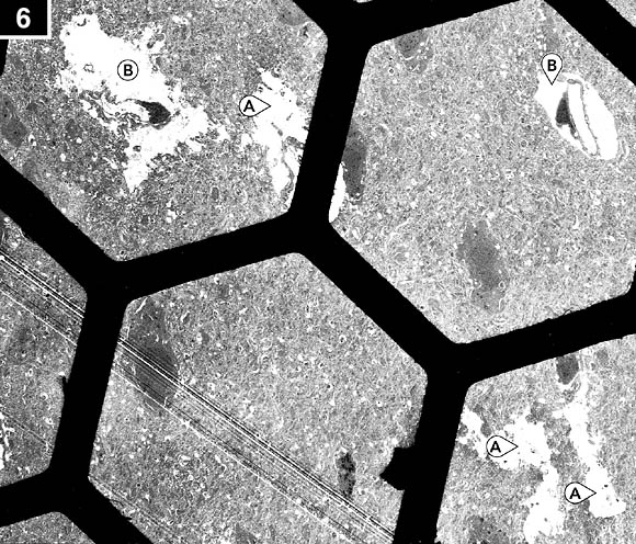
Simplified protocol. The brain was glycerolized to a lower level (4M) at a faster rate (700 mM/minute) before being frozen to –77°C for one week, thawed and reperfused with fixative. This simplified perfusion is NOT used by Bio-Preservation on its human patients but is similar to practices which were typical in cryonics up to the 1980s.
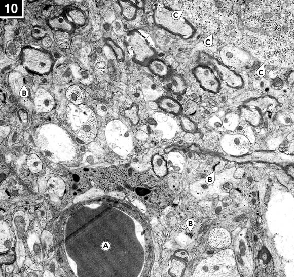
No perfusion or freezing. To provide control data, these samples were taken from anesthetized dogs that were perfused with fixative, not glycerol, and were not cooled at all.

1. BioPreservation protocol. Typical appearance of gray matter at 6700x magnification. Note intact capillary endothelial cells (A) and particles of carbon (B) in the capillary lumens. The overall appearance of the neuropil and of the axons and neurons is excellent.

2. BioPreservation protocol. Neuropil in gray matter from the hippocampus at 35,500x magnification. Architecture of intracellular components, synapses (A), and neuronal membrane integrity are excellent.

3. BioPreservation protocol. A synapse in gray matter from the hippocampus at 40,200x magnification. The presynaptic junction contains small packets of neurotransmitter (A) visible as granules. Note the overall crisp appearance of both the synaptic membranes and adjacent structures of the neuropil. This degree of preservation at the synaptic level was uniformly observed in all samples examined.

4. BioPreservation protocol. Gray matter from the hippocampus at 6700x magnification showing two of many synapses (A) and some defects in myelin (B). The axoplasm is intact, with good internal structure and overall high-quality appearance of the neuropil (weave of brain connections) by comparison with similar samples using the simplified protocol. The heavy, wiggly lines across the center of the picture are myelinated axons.

5. BioPreservation protocol. Closeup of the same area of gray matter at 40,200x magnification. Note that while myelin is injured (A), the axoplasm within the myelin exhibits excellent structural preservation and the membranous structure of the neuropil is intact as are intracellular organelle membranes. A mitochondrion (B) is visible.

6. Simplified protocol. Hippocampus at 2000x magnification. The black hexagons are the copper grid on which the specimen rests in the electron microscope. Note the frequency of large ice holes or “tears” (A) in the neuropil and the uniform presence of pericapillary ice holes (B).

7. Simplified protocol. Gray matter from the hippocampus showing very poor reperfusion. A large pericapillary ice hole almost completely severs the capillary from the neuropil (A). The nucleus is denuded of cytoplasm (B) and the endothelial cell has lost its plasma membrane with only bits of cytoplasm clinging to the basement membrane (C). There are also ice holes (D) and a generalized loss of membranous structure with apparent reorganization into vacuoules. This kind of injury is typical of 4M brains prepared in this way.

8. Simplified protocol. A badly damaged capillary at 9700x magnification. Two large tears (A) are visible, with debris scattered in the capillary (B). There is a naked nucleus (C) with no nuclear membrane, from a lysed cell.

9. No perfusion or freezing. Gray matter from the hippocampus at 40,000x magnification. Synapses are present (A) as are mitochondria (B). Interestingly, there are defects in the myelin of several axons even in this specimen from a control dog that was not frozen (C).

10. No perfusion or freezing. Gray matter at 15,000x magnification. Note the red cell (A) in the capillary. Lack of dehydration is evidenced by more fine structure in the neuropil and within the axons, lack of shrinkage of the axoplasm, and a more homogeneous character of the myelin. Many synapses are present (B). The boundary of a neuron is visible (C).

11. BioPreservation protocol. White matter from the corpus collosum at 6700x magnification. Note the excellent preservation of the capillary (A) and its endothelial cell plasma membranes. The nucleus (B) shows typical loss or reorganization of nucleoplasm; this is seen more frequently in frozen-thawed brains than in brains just perfused with glycerol and fixed without freezing. Several axons (C) exhibit typical skrinkage of axoplasm and alteration in myelin structure. The increase in free space between axons and other structures is the result of glycerol-induced dehydration.

12. BioPreservation Protocol. The injury visible here was seen rarely in these samples. Note the presence of what appear to be pericapillary ice holes (A). The endothelial cell membranes of the capillary appear indistinct in several places (B) and there seem to be a few small “blebs” or vesicles of cell membrane material in the capillary lumen. The dark black specks (C) near the endothelial cells are carbon particles which were present in the fixative. Note also the peculiar pattern of injury to the cell nucleus (D) wherein it appears that nuclear material has been lost or rearranged. The nucleolus is also very shrunken.
